Home
/ Back Muscles Anatomy Ct - Transversospinales Physiopedia _ Schau dir angebote von top brands auf ebay an.
Back Muscles Anatomy Ct - Transversospinales Physiopedia _ Schau dir angebote von top brands auf ebay an.
Back Muscles Anatomy Ct - Transversospinales Physiopedia _ Schau dir angebote von top brands auf ebay an.. The extrinsic back muscles are located in the back, but act to produce movements of the shoulder and assist respiration. The intrinsic back muscles are found deeper to the extrinsic muscles, separated from them by the thoracolumbar fascia. It allows to differentiate the vertebrae, the nervous system, the intervertebral discs and the zygapophyseal joints. The muscles of the chest and upper back occupy the thoracic region of the body inferior to the neck and superior to the abdominal region and include the muscles of the shoulders. Schau dir angebote von top brands auf ebay an.
The following slides are from wikiradiography (wetpaint) here. The muscles of the chest and upper back occupy the thoracic region of the body inferior to the neck and superior to the abdominal region and include the muscles of the shoulders. Filed under anatomy, ct, head and neck. The muscles of the back can be arranged into 3 categories based on their location: The neck consists of seven cervical vertebrae, the building blocks of the spine.
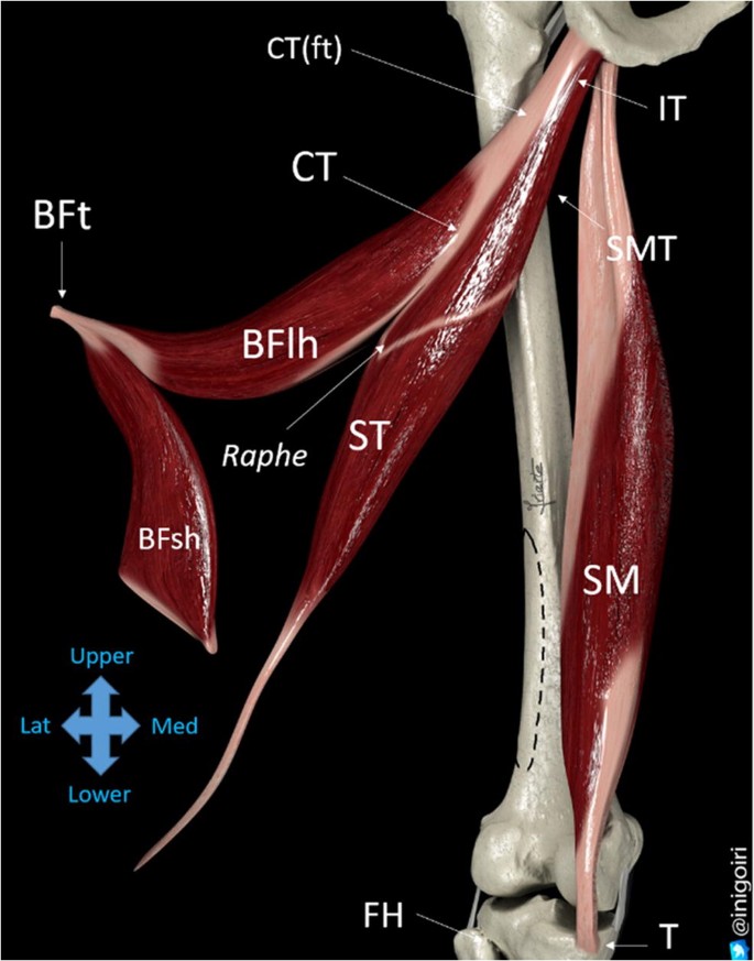
Sonographic Landmarks In Hamstring Muscles Springerlink from media.springernature.com Über 7 millionen englischsprachige bücher. Folge deiner leidenschaft bei ebay! On this page, you'll learn about each of these muscles, their locations and functional anatomy. Computed tomography (ct) is a proven method for evaluating the lumbar spine. Other muscles are small and cover much less space. Schau dir angebote von top brands auf ebay an. Muscles of the lumbar spine. Anatomy of back muscles your back consists of three distinct layers of muscles, namely the superficial layer, the intermediate layer, and the deep layer.
Computed tomography (ct) is a proven method for evaluating the lumbar spine.
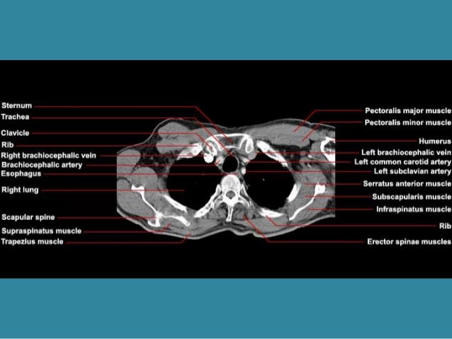
Able to move the upper limb as they originate at the vertebral column and insert onto either the clavicle, scapula or humerus. The chest or thorax is the region between the neck and diaphragm that encloses organs, such as the heart, lungs, esophagus, trachea, and thoracic diaphragm. Posted by radiologypics ⋅ march 21, 2013 ⋅ 1 comment. Superficial back muscles, intermediate back muscles and intrinsic back muscles.the intrinsic muscles are named as such. The muscles of the back can be arranged into 3 categories based on their location: Superficial back muscles, intermediate back muscles and intrinsic back muscles.the intrinsic muscles are named as such because their embryological development begins in the back, oppose to the superficial and intermediate back muscles which develop elsewhere and are therefore classed as extrinsic muscles. Other muscles are small and cover much less space. In the upper back region, the trapezius, rhomboid major, and levator scapulae muscles anchor the scapula and clavicle to the spines of several vertebrae and the occipital bone of the skull. The images are available in the three planes, axial, sagittal. This mri chest (thorax) axial cross sectional anatomy tool is absolutely free to use. Each block is separated by a disc that sits in between and each vertebra has a facet joint on either side. Filed under anatomy, ct, head and neck. All about the back muscles the back anatomy includes the latissimus dorsi, trapezius, erector spinae, rhomboid, and the teres major.
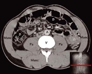
Abdominal_anatomy_back_view 2/3 abdominal anatomy back view. Back pain, and ct was ordered after aaa was identified on radiography. To perform clinical clinical orthopedic manual therapy to the lumbar spine. On this page, you'll learn about each of these muscles, their locations and functional anatomy. The back muscles are divided into two large groups:
Anatomy Of Chest from image.slidesharecdn.com Muscle anatomy gluteus 12 photos of the muscle anatomy gluteus gluteus muscle anatomy ct, gluteus muscle anatomy mri, human muscle anatomy gluteus maximus, muscle anatomy gluteus, muscle anatomy of gluteal, human muscles, gluteus muscle anatomy ct, gluteus muscle anatomy mri, human muscle anatomy gluteus. Abdominal_anatomy_back_view 2/3 abdominal anatomy back view. This blog post article is an overview of the muscles of the lumbar spine of the trunk. Normal radiographic anatomy by dr. The images are available in the three planes, axial, sagittal. Über 7 millionen englischsprachige bücher. Related posts of muscles shoulder and back muscle anatomy lower extremity. Iliocostalis subgroup is the most lateral longissimus subgroup is between iliocostalis and spinalis
All about the back muscles the back anatomy includes the latissimus dorsi, trapezius, erector spinae, rhomboid, and the teres major.
Normal radiographic anatomy by dr. The muscles of the back can be arranged into 3 categories based on their location: Attached to the pelvis are muscles of the buttocks, the lower back, and the thighs. Includes latissimus dorsi, the trapezius, levator scapulae and the rhomboids. This group is made of three subgroups, with the group divisions occurring by location. The superficial back muscles are situated underneath the skin and superficial fascia. The extrinsic back muscles are located in the back, but act to produce movements of the shoulder and assist respiration. Abdominal_anatomy_back_view 2/3 abdominal anatomy back view. The back muscles are divided into two large groups: Msk_lower limb by angelo gambino. Back pain, and ct was ordered after aaa was identified on radiography. The intrinsic back muscles are found deeper to the extrinsic muscles, separated from them by the thoracolumbar fascia. The images are available in the three planes, axial, sagittal.
Anatomy of back muscles your back consists of three distinct layers of muscles, namely the superficial layer, the intermediate layer, and the deep layer. The muscles of the chest and upper back occupy the thoracic region of the body inferior to the neck and superior to the abdominal region and include the muscles of the shoulders. The back muscles are anatomically layered into superficial (extrinsic) and deep (intrinsic) muscles. Anatomia by dr césar reyes. These muscles, including the gluteus maximus and the hamstrings, extend the thigh at the hip in support of the body's weight and propulsion.
Abdominal Ct Anatomy Radiology Key from radiologykey.com Muscle anatomy gluteus 12 photos of the muscle anatomy gluteus gluteus muscle anatomy ct, gluteus muscle anatomy mri, human muscle anatomy gluteus maximus, muscle anatomy gluteus, muscle anatomy of gluteal, human muscles, gluteus muscle anatomy ct, gluteus muscle anatomy mri, human muscle anatomy gluteus. Über 7 millionen englischsprachige bücher. Each block is separated by a disc that sits in between and each vertebra has a facet joint on either side. Includes latissimus dorsi, the trapezius, levator scapulae and the rhomboids. Lower back pain is a pervasive symptom. Anatomia by dr césar reyes. The muscles of the chest and upper back occupy the thoracic region of the body inferior to the neck and superior to the abdominal region and include the muscles of the shoulders. Filed under anatomy, ct, head and neck.
Back pain, and ct was ordered after aaa was identified on radiography.
Muscles of the lumbar spine. (2017, elsevier) should be consulted. Related posts of muscles shoulder and back muscle anatomy lower extremity. The erector spinae group is the intermediate layer of the intrinsic muscles of the back. Includes latissimus dorsi, the trapezius, levator scapulae and the rhomboids. The muscles of the back can be arranged into 3 categories based on their location: This mri chest (thorax) axial cross sectional anatomy tool is absolutely free to use. The seventh cervical vertebra, referred to as c7, meets the first of 12 thoracic vertebrae t1 at the base of the neck, a. Lower back pain is a pervasive symptom. Vertebrae_anatomy_ct 2/3 vertebrae anatomy ct epub vertebrae anatomy ct vertebrae anatomy ct bones. Normal radiographic anatomy by dr. The neck consists of seven cervical vertebrae, the building blocks of the spine. Each block is separated by a disc that sits in between and each vertebra has a facet joint on either side.
The following slides are from wikiradiography (wetpaint) here back muscles anatomy. Posted by radiologypics ⋅ march 21, 2013 ⋅ 1 comment.